Yuan Zhang1,
Hui Zhang1,
Yitong Ma2,
Yining Yang2,
Miaomiao Ma1,
Xiaoli Zhu1,
Li Wang1 ![]()
For correspondence:- Li Wang Email: wanglishz@163.com Tel:+869932858754
Received: 21 October 2015 Accepted: 13 March 2016 Published: 30 April 2016
Citation: Zhang Y, Zhang H, Ma Y, Yang Y, Ma M, Zhu X, et al. Effect of β-adrenoceptor on cardiac fibrosis in rat cardiac fibroblast cells and its potential mechanism. Trop J Pharm Res 2016; 15(4):717-722 doi: 10.4314/tjpr.v15i4.7
© 2016 The authors.
This is an Open Access article that uses a funding model which does not charge readers or their institutions for access and distributed under the terms of the Creative Commons Attribution License (http://creativecommons.org/licenses/by/4.0) and the Budapest Open Access Initiative (http://www.budapestopenaccessinitiative.org/read), which permit unrestricted use, distribution, and reproduction in any medium, provided the original work is properly credited..
Purpose: To investigate the effect of β3-adrenoceptors (β3-AR) up-regulation on fibrosis in cardiac fibroblast cells in rats and its potential mechanism.
Methods: Cardiac fibroblast cells (CFB) were isolated and identified from rats’ hearts. The β3-ARup-regulated cardiac fibroblast cells were constructed by lentiviral transfection technology. Thereafter, Ang II was used to induce fibrosis in cardiac fibroblast cells, and subsequently, Western blot assay was performed to investigate fibrosis related marker proteins (TGF-β, Smad-2, p-Smad-2, Col-I and Col-III) in cardiac fibroblast cells.
Results: β3-AR up-regulated cardiac fibroblast cells were successfully constructed. Furthermore, the results show that up-regulation of β3-AR increased the ex
Conclusion: The results suggest that up-regulation of β3-AR aggravates fibrosis of cardiac fibroblast cells. In other words, inhibition of β3-AR ex
Introduction
Cardiovascular disease is one of the leading causes of death all over the world [1]. Cardiac fibrosis is characterized by excessive deposition of collagenous fiber in the extracellular matrix (ECM) and disproportionate increase and disordered arrangement of myocardial collagen [2-4]. Increasing evidence demonstrated that cardiac fibrosis (CF) is a central factor for the development of several serious cardiovascular diseases, including cardiac failure, myocardial infarction and cardiac hypertrophy, etc [2,4-6]. Therefore, discovering more novel reliable cardiac fibrosis interventions might be a feasible approach to cure various serious heart diseases.
Previous investigations revealed that β3-Adrenoceptors (β3-AR) is lowly expressed in normal human and animal ventricle myocardium, and the activation of β3-AR could affect myocardial contraction and plays an important role in the pathogenesis of cardiac dysfunction [7-9]. However, so far, there is no reported work regarding the effects of β3-AR on cardiac fibrosis, thus our present study is designed to investigate the potential of β3-AR on fibrosis in cardiac fibroblast cell and its potential mechanism. In our present work, β3-AR over-regulated cardiac fibroblast cells were constructed by lentiviral transfection. Then, Ang II was used to induce fibrosis in the cardiac fibroblast cell, and subsequently western blot assay was performed to investigate the fibrosis related marker proteins in the cardiac fibroblast cells.
Methods
Animals
SD sucking rats (1 - 3 days), purchased from the experimental animal center of the first affiliated hospital of Xinjiang Medical University (Urumqi, China), were used in the present investigation. The protocol for the use of animals was according to the guidelines of animal experiments from the ethical committee for animal research of the First Affiliated Hospital of the Medical College, Shihezi University (ethical approval o.: S2013-04-02).
Reagents and chemicals
The resulting recombinant lenti viruses (Objective gene sequence number: NM_013108.2) were purchased from Cyagen Biosciences Inc., (Guangzhou, China). DMEM medium and fetal bovine serum (FBS) were purchased from Gibco Co. (Shanghai, China); β3-ARprimary antibody was purchased from Santcruz Co. (Japan), TGF-β and Collagen-III (Col-III) primary antibodies were purchased from Protein tech Co. (USA), Smad-2/3, Phospho-Smad-2,vimentin, horseradish- peroxidase-conjugated secondary antibodies and GAPDH primary antibodies were purchased from Cell Signaling Co. (USA), Col-III primary antibody was purchased from Abcam (UK); BCA protein assay kit was purchased from Beyotime (Jiangsu, China). All other regents and chemical used in our present study were analytical grade.
Primary culture of rat ventricular cardiac fibroblasts
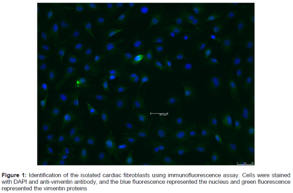
Rat cardiac fibroblasts were isolated and cultured according to previously described method [4]. Briefly, hearts of the rats were removed and chopped into 1 mm3 pieces, then digested in 0.1 % type II collagenase and 0.25 % trypsin. Subsequently, the cell suspension was centrifuged to collect the cell pellet, then the cell pellet was re-suspended with DMEM containing 10 % FBS. After 1 h incubation in culture flask, the attached cells were further cultured at 37 °C in a humidified and 5 % CO2 atmosphere. The isolated cardiac fibroblasts were identified by using the immunofluorescence assay with vimentin protein, and our results showed that the vimentin protein positive cells accounted for more than 95 % (). The second to fourth passages of cardiac fibroblasts were used for our research.
Preparation of β3-AR up-regulated cardiac fibroblast cells
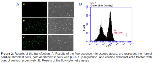
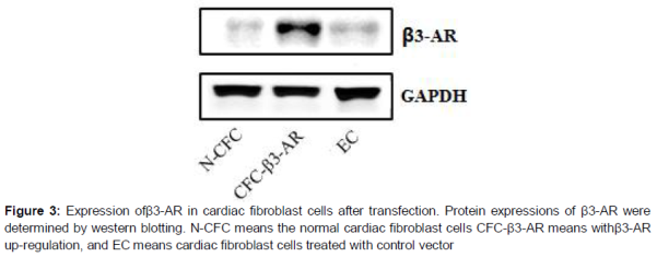
The β3-AR gene siRNA vector recombinant plasmid was constructed using RNA interference technique. Then the resulting recombinant lentiviruses were used to infect cardiac fibroblast cells to up-regulate the β3-AR. The expression of β3-AR in normal cardiac fibroblast cells (N-CFC), cardiac fibroblasts cells with β3-AR up-regulation (CFC-β3-AR), and cardiac fibroblast cells treated with control vector (EC) were detected using western blotting and fluorescence microscope observation. In addition, flow cytometry assay was performed to determine the transfection rate.
Western blot
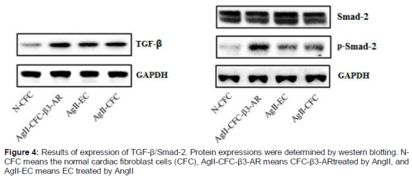
After lentiviral transfection, the cardiac fibroblast cells were induced to fibrosis by treating with Ang II (10-6M) for 24 h [4,10]. Then, the cardiac fibroblast cells were cultured for extraction of total proteins. Subsequently, the protein concentration was determined using the BCA Protein Assay kit. Then equal amounts of proteins (35 μg) were separated by SDS/PAGE, blotted on PVDF membrane and probed with various primary antibodies (including β3-AR, TGF-β, Smad-2, Col-I and Col-III), followed by incubation with the secondary antibodies and chemiluminescence detection. To normalize for protein loading, antibodies directed against GAPDH were used.
Statistical analysis
Data are expressed as mean ± SD of three independent experiments. Statistical analysis was carried out using SPSS 15.0 software package and one way analysis of variance (ANOVA) with Dunnett’s test to compare the means between two groups with a significance level of p < 0.05.
Results
Expression of β3-AR in cardiac fibroblast cells after transfection
As can be seen from our results of fluorescence microscope assay which is shown in A, after transfection, the β3-AR proteins were significantly up-regulated in the cardiac fibroblast cells, compared with both the N-CFC and EC groups, and the results of the flow cytometry assay showed that the transfection rate was nearly 70 %. Furthermore, the western blot results also indicated that β3-AR proteins expressions increased after transfection (). These results suggest that the cardiac fibroblast cells with β3-AR up-regulation were constructed successfully.
Effect of β3-AR up-regulation
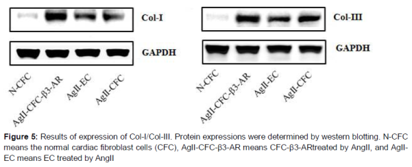
After being induced by Ang II for 24 h, TGF-β, Smad-2, Col-I and Col-III expressions in cardiac fibroblast cells were determined by western blotting assay. As can be seen from , our results showed that no change was observed for Smad-2 expressions among all the test groups. However, it is interesting that TGF-βand p-Smad-2 were significantly up-regulated in β3-AR up-regulated cardiac fibroblast cells, compared with the N-CFC, AgII-EC and Ag II-CFC. Furthermore, the collagen types I and III proteins (Col-I and III) were also determined in our present study, and our results revealed that both Col-I and Col-III were up-regulated in cardiac fibroblast cells by up regulation of the β3-AR (). These results indicate that up-regulation of β3-AR could promote cardiac fibrosis in cardiac fibroblast cells.
Discussion
Tissue fibrosis can result in certain serious diseases for which effective therapy is lacking [11]. For the heart, cardiac fibrosis damages cardiac contraction and causes cardiac dysfunction [2]. Therefore, inhibition of cardiac fibrosis would be beneficial for improving some serious cardiac diseases. To the best of our knowledge, the present work is the first reported study on the effect of β3-AR on the development of cardiac fibrosis, which has significant reference value for the future treatment of cardiac diseases in the clinic.
β3-AR is commonly expressed at low levels in cardiac tissues, and previous research revealed that β3-AR would be up-regulated in patients with dilated cardiomyopathy [8]. Therefore, we guessed that β3-AR might play an important role in the pathogenesis of cardiac fibrosis. In our study, the lentivirus transfection technology was used to construct the β3-AR over-expressed cardiac fibroblast cells. Previous reports have demonstrated that Ang II is a well-established factor related to many cardiac diseases and could induce the development of cardiac fibrosis [12,13]. Thus, Ang II induced cardiac fibrosis strategy is a commonly used experimental model both in vivo and in vitro [4,14,15]. In our present study, the fibrotic cardiac fibroblasts cells were induced successfully with Ang II. TGF-β/Smads is a well-known pathway involved in multi-organ tissue fibrosis via inducing endothelial-mesenchymal transition (EndMT) and vascular remodeling, etc. [16,17]. TGF-β acts by activating Smads signaling pathway via phosphorylating the Smad-2/3 proteins. The p-Smads subsequently translocates from the cytoplasm into the nucleus to regulate other transcription factors [18,19]. Therefore, the expressions of TGF-β/Smads could reflect the extent of cardiac fibrosis. Furthermore, collagen is the most abundant protein in animal tissues and also the major component in the interstitial extracellular matrix. Previous researches have revealed that Col-I and Col-III would be over-expressed in patients with cardiac fibrosis, and Col-I/III could be considered as the biomarkers of cardiac fibrosis [20,21]. In our present investigation, up-regulation of β3-AR increased the expressions of Col- I and Col- III. Both our results mentioned above suggested that up-regulation of β3-AR aggravated the fibrosis of cardiac fibroblast cells. In other words, inhibition of β3-AR expression in cardiac tissues would be beneficial for treating cardiac fibrosis and its related cardiac diseases.
Conclusion
The findings of this work demonstrate that up-regulation of β3-AR up-regulates the expressions of TGF-β, Smad-2, Col-I and Col-III in rat cardiac fibroblasts cells, thus suggesting that up-regulation of β3-AR aggravates cardiac fibrosis. Thus, this present study also indicating that down-regulating β3-AR might be a useful strategy for treating cardiac fibrosis in clinic.
Declarations
Acknowledgement
References
Archives


News Updates